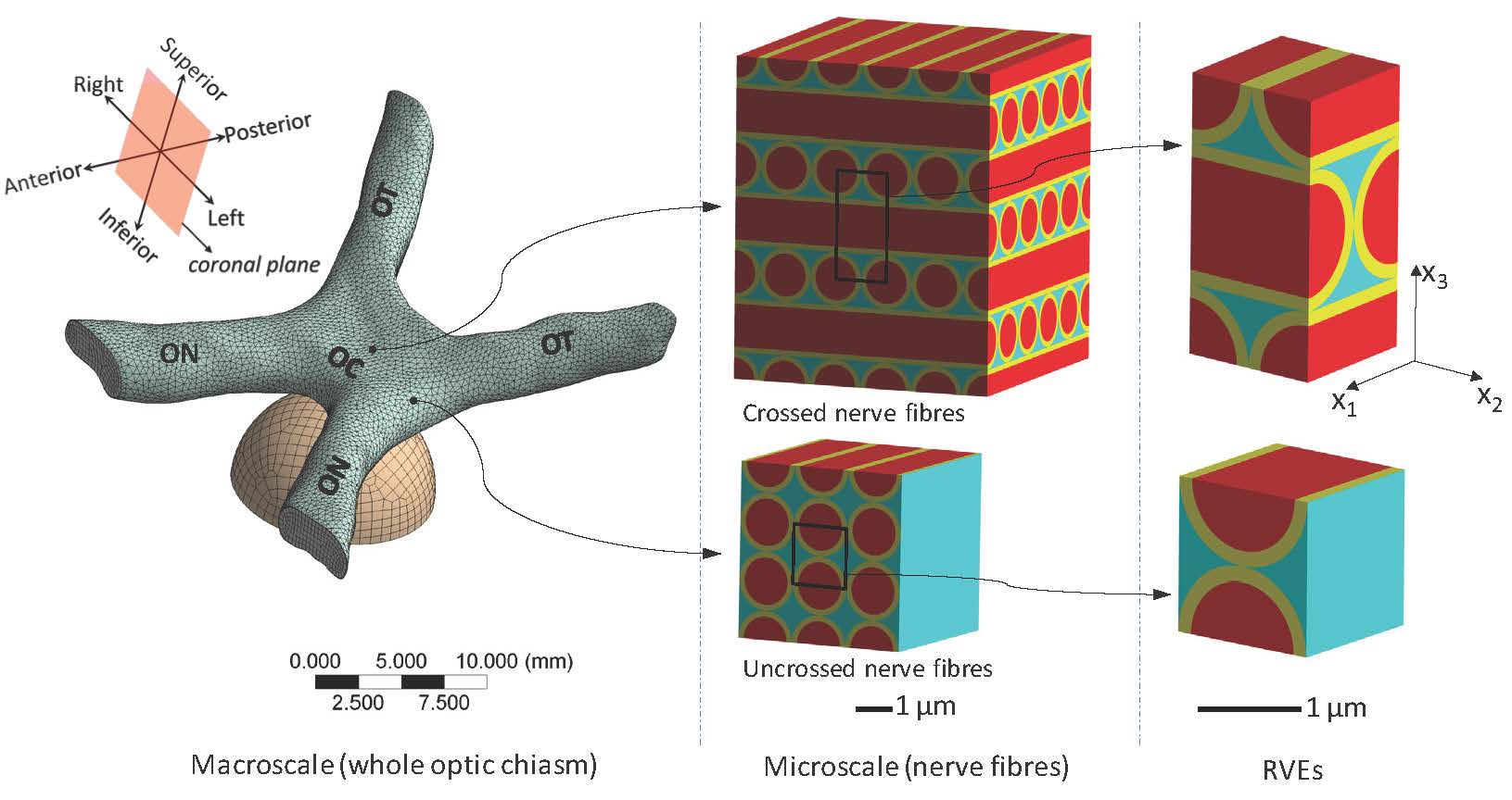
UNSW Canberra is investigating the role mechanical compression of the optic chiasm plays in visual afflictions (Wang et al. 2014a). The nerves carrying optical information from both eyes all pass through this small soft-tissue structure inside the skull. This research is being performed in collaboration with Canberra Hospital, the ANU Medical School and Queens University, Belfast. The work uses high-resolution histology of chiasm anatomy to inform multi-scale numerical models of the biomechanics of chiasmal compression (Wang et al. 2014b). These numerical models are being validated using three-dimensional imaging of chiasm compression experiments performed in vitro. Increased fidelity of these models requires more precise knowledge of the three-dimensional axonal routing within the chiasm and the anatomy of the surrounding soft tissue. Current work is using high-resolution imaging of the chiasm to map the axonal pathways for inclusion in the biomechanical models (Jain et al. 2015).
Image shows The macroscopic finite-element model of the optic chiasm and tumour and the microscopic representative volume element (RVE models) of the packed nerve axons. (OC: optic chiasm; ON: optic nerve; OT: optic tract.).